Contents
Overview
In this exercise, we will:
- Download a PDB structure of a mutated protein. In its native state, the protein is glycosylated, and it does not perform its functions properly without at least some of the glycosylation.
- Use the text editor and the utilities at GLYCAM-Web to change the mutated residues back to the natural ones.
- Use GLYCAM-Web to glycosylate the un-mutated protein.
In the case of the structure 1BUY, an NMR structure determined in aqueous solution, it was necessary to cause mutations in the protein before a 3D structure could be obtained. Specifically, to improve solubility, the three N-glycosylation sites were mutated from asparagine (ASN) to lysine (LYS). You can read the paper (reference available at the RCSB site) to learn this, but the PDB file itself contains hints regarding the changes that were made. We will open the PDB file to see these hints and edit it to change the three LYS residues back to ASN.
Download 1BUY.pdb
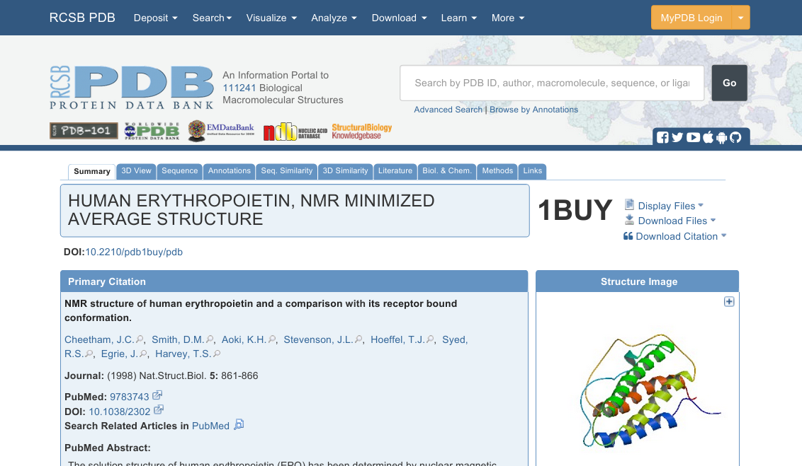
Direct your web browser to rcsb.org. In the search box at right near the top, type “1BUY” and click Go. You should land on a page similar to the one shown below. Spend a moment or two browsing around on the page to get a feel for the information that is available. When you are ready, click on Download Files (to the right of the big 1BUY) and download the PDB File (Text).
Inspect the PDB File
Open the pdb file using your text editor. The first hints about the changes made to the protein occur on lines 7 and 8, where it is declared to be engineered and to have been mutated.
Find the locations of the mutations
Now, scroll down to lines 209-211. On SEQADV lines, any conflicts between the protein sequence in the PDB file and the sequence reported in another database are noted. The PDB file doesn’t attempt to declare that one is “correct” and the other is “wrong”. It merely notes on these lines the differences. These specific lines say that in structure 1BUY, the three LYS residues in chain A numbered 24, 38, and 83 differ from another database of protein structures, and that in the other database, those residues are asparagines. Each line also includes the comment “ENGINEERED”, which is another significant hint. Of course, for research, you should always read the original paper to be certain that you have fully understood information found in a PDB file. In this case, these are the three mutagenesis locations.
For more information on the SEQADV card format, see:
http://www.wwpdb.org/documentation/file-format-content/format33/sect3.html#SEQADV
Now all we need to do is change the lysines back to asparagines. For computational molecular modelers this is a common and relatively simple task.
Change the file to reverse the mutations
This procedure is greatly simplified by the fact that amino acid structures differ only in the side chains. So, we will delete the side chains from the LYS residues, and change the residue names for the remaining atoms to ASN. In the next step, we will let the Glycoprotein Builder rebuild the side chains. Then, we will save the new file with the ASN residues restored. Later, we will glycosylate it.
- Scroll down to residue LYS 24. It starts on line 618.
- Delete all the atoms except the first four (N, CA, C, and O).
- Change LYS to ASN on the four remaining lines of the LYS residue.
- Caution! You must perfectly preserve the spacing in this file. Make sure that the columns of characters are still aligned properly after you make the change. This is very important.
- Do not be concerned about the atom serials (atom numbers). They do not need to be sequential.
- Repeat this procedure on LYS 38 and 83.
- Save the file with a new name, such as 1BUY_edited.pdb (1BUY_edited.txt is also OK if that is simpler).
Rebuild your reverse-engineered 1BUY
- Go to glycam.org/gp and upload your edited file.
- Click Continue at the Preprocessor stage.
- Do not add glycans yet! Click Download current structure without minimization.
- We need the builder to build the ASN residues back in so that they can be properly glycosylated.
- Download the Amber style with H If desired, change the name from structure_AMBER.pdb.
- Optionally: open the file in VMD or your text editor to confirm that the residues have been rebuilt to full ASNs.
Glycosylate your rebuilt EPO
- Go back to glycam.org/gp and upload the rebuilt PDB file. Fill in the other boxes as you please.
- Choose a High Mannose structure from the Oligosaccharide library and add it to the three rebuilt ASN residues.
- The image below was made using Man9GlcNAc2. If you choose a smaller one, it will take less time to process, but “resid 200” will probably be some number other than 200 for you.
- Click Continue and Review Current Glycans. Confirm the proper addition of the glycans.
- Now, use the interactive carbohydrate builder to build DGalNAcb1-OH and click Done.
- Add the GalNAc to O-linking site 126 (you will need to SHOW ALL), and click Continue.
- Review Current Glycans to ensure you have built what you intended, and, when ready, click Download current structure without minimization.
- Again, the wait page might take a few seconds. If you try on your own later, minimization will take ~2 min.
- Download the Amber style with H file (or the zip archive) and visualize it online or using VMD.
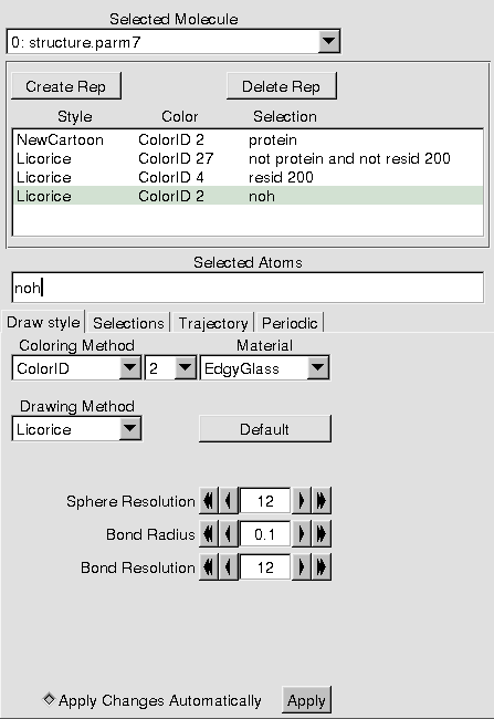
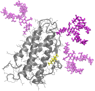
Use VMD to display the glycosylated structure
Here are instructions for the image below. The graphical representations are as shown in the adjacent image.
- To achieve the white background, go Main -> Graphics -> Colors -> Display -> Background -> White.
- The bond radius for the ‘noh’ rep is 0.1, and for the glycans, it is 0.4.
- Note that, depending on the nature of the glycans you added, the resid of the GalNAc might not be 200. Use the mouse to label one of the atoms so that you can determine its number.
- I rendered the image specially. Depending on the system you are using, this might not be practical because either you need extra software or because your computer is slow. To try, go File -> Render. In the first pull-down, choose Tachyon. Change the filename if you like (perhaps glycosylated_EPO) and click Start Rendering. Note that on Windows, you will need to have set a Working Directory (see Introduction to VMD).
If there is extra time
Glycosylate another protein (e.g., 1RBB). Or, play around with different representations of EPO. Search the PDB for “glycoprotein” and see what you find.
